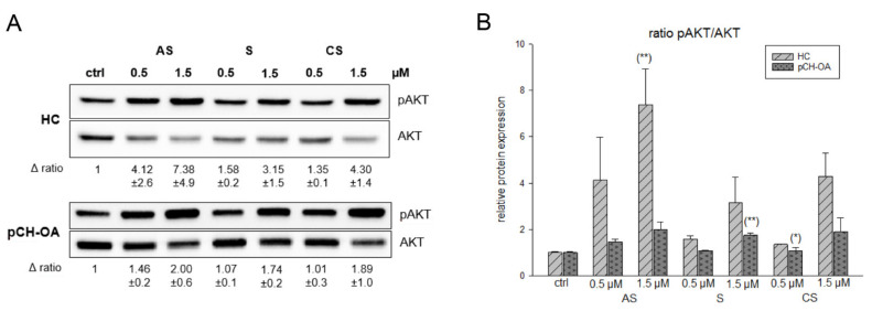
Figure 3.
Shikonin derivatives induced an activation of AKT phosphorylation. (A) The AKT protein phosphorylation was evaluated by immunoblotting under untreated control conditions (ctrl) and in the presence of 0.5 µM and 1.5 µM acetylshikonin (AS), shikonin (S), and cyclopropylshikonin (CS) for 1 h in HC and IL-1β-stimulated pCH-OA cells. AKT was used as loading control. Δ ratio represents the fold change of pAKT/AKT normalized to controls (mean ± SD, n = 3). (B) shows the densiometric evaluation of all experiments in HC (light grey striped) and pCH-OA (dark grey dotted) cells. Statistical significances are defined as follows: * p < 0.05; ** p < 0.01.

