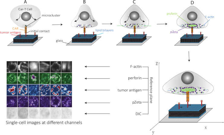
Fig 2. The model shows the process of perforin and pZeta cluster formation, and accumulation of F-actin formation after the initial contact of the CAR-T and planar lipid bilayer.
(A) At the initial contact of the CAR with the tumor antigen, micro clusters are formed around the receptor, and the cell starts to spread. (B) The cell spread, and multiple microclusters form. (C) After the cell spread, F-actin polymerizes at the cell periphery. The perforin and pZeta are transported toward the cell center along with F-actin. (D) Perforin and pZeta populate the actin-sparse center and form a cluster. In the experiment, we labeled the different substances with different colors, and different channels of images were obtained using different lasers. We use six single-cell samples in five channels using the best Z position.

