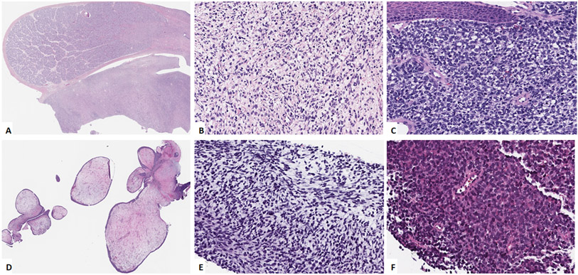
Figure 1: Morphologic spectrum of NRAS-mutated RMS.
A, B. (case 2, 2/M) Paratesticular tumor (A) showing primitive appearing cells in a loose stroma (B). C. Soft palate lesion (case 3, 2/M) composed of undifferentiated cells with round to irregular nuclei arranged in sheets, involving submucosa. D. Vaginal tumor (case 5, 3/F) showing botryoid morphology with polypoid nodulear growth. E. Jaw lesion (case 6, 5/M) showing primitive ovoid to spindle cells in a myxoid background. F. Calf tumor (case 11, 62/M) showing undifferentiated small cells arranged in sheets.

