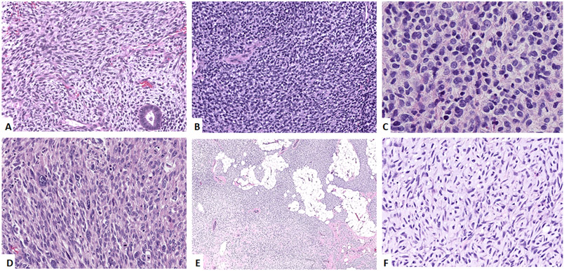
Figure 2: Morphologic spectrum of KRAS- (A-C), HRAS- (D) and BRAF-mutated (E, F) ERMS.
A. Uterine tumor (case 19, 44/F) showing primitive-appearing spindle cells involving the endometrium. B. Vaginal tumor (case 16, 1/F) showing undifferentiated cells with round nuclei in sheets. C. Paratesticular mass (case 22, 20/M) showing sheets of undifferentiated round cells. D. Thigh lesion (case 25, 50/M) showing a sarcomatoid morphology with spindle to pleomorphic cells in a fascicular pattern. E, F Abdominal tumor (case 26, 4/M) showing a cellular tumor infiltrating adipose tissue. Higher power image (F) showing primitive spindle cells in a collagenous to myxoid stroma.

