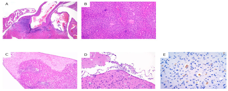
Figure 3.
Histopathological analysis of trypanosome-infected mice at 28 dpi. (A) necrotizing arteritis in the heart (hematoxylin and eosin (HE) staining) in group III; (B) vasculitis and perivasculitis in the liver (HE staining) in group III; (C) focal necrosis in the liver (HE staining) in group IV; (D) meningoencephalitis in the brain (HE staining) in group IV; and (E). trypanosomes in the brain (immunohistochemical staining for trypanosomes, brown) in group IV.

