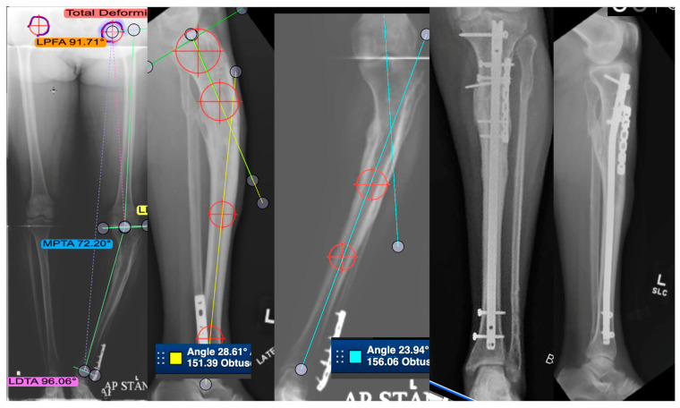
Figure 4.
This is a case of a 53-year-old patient with post-traumatic tibial deformity complaining of knee and ankle pain. The deformity was assessed on long-leg standing alignment views, compared to the other tibia in AP and lateral views. CT scan revealed very little rotational deformity. An osteotomy was planned at the center of rotation and angulation (CORA), which was close to the same location on both AP and lateral. This is a single-plane deformity, which has been measured on both the AP and lateral images. The right two images show the patient post-transverse opening-wedge osteotomy fixed with an intramedullary nail and a plate. Healed in good alignment.

