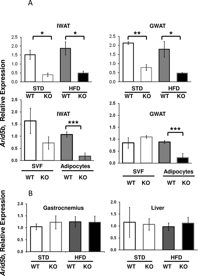
Figure 1. Arid5b Expression in Cells and Tissues of WT and FSKO mice.
A. Arid5b expression in WAT, adipocytes and SVF. IWAT, and GWAT, were isolated from WT and FSKO mice that had been maintained on STD or HFD. Fat tissue was digested with collagenase, filtered and centrifuged as described in Methods, and RNA was extracted from both floating adipocytes (solid bars) and the stromovascular fraction (SVF-open bars). B. Arid5b expression in liver and gastrocnemius muscle was only isolated from WT and FSKO mice on HFD. Arid5b expression was determined using RT-PCR as described in Methods. Values are means ± SE of tissues from 6 animals in each group. *P < 0.05, **P < 0.01, ***P < 0.001.

