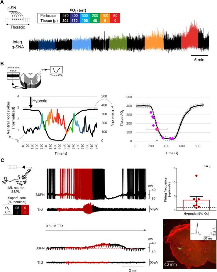
Fig. 5. Neonatal sympathetic responses to hypoxia in perfused in situ thoracic spinal cord and en bloc transverse thoracic spinal cord section preparations.
(A) Recording of neonatal (P2) g-SNA in response to hypoxia in a single perfused thoracic spinal cord in situ preparation (schematic in top left corner). Reducing perfusate PO2 increases g-SN activity. Box: Color-coding illustrates perfusate PO2 in torr. Amplitude and/or area under the curve indicates activity level. (B) IML tissue PO2 measurement from Clark-style microelectrode with tip 100 μm below tissue surface with contralateral ventral nerve recording from en bloc transverse thoracic spinal cord section preparation (schematic in top left corner). As tissue PO2 decreases, ventral nerve activity increases (i.e., mounted responses exceeding four SDs of baseline activity) in 8 of 14 preparations. PO2 threshold response (as defined above) occurred at 140 ± 121 torr (n = 8). (C) Individual IML SSPN (whole cell) and ventral root nerve activity (T2) in en bloc preparations. Firing frequency increased (as defined above) in 8 of 11 preparations when aCSF equilibrated with 0% O2 was delivered to the dish (giving a dish nadir ~13% FO2). During the 1-min peak response, firing rate increased by 3.67 ± 1.32 spikes/s (n = 8). In TTX, hypoxia depolarized neurons by 3.1 ± 0.98 mV (repeated-measures one-way ANOVA; baseline versus hypoxia, P = 0.03; hypoxia versus washout, P = 0.01; n = 10) but had no effect on membrane resistance (1049 ± 29 megohms; repeated-measures one-way ANOVA, P = 0.5; n = 10). Neurons were identified by backfiring the ventral root and filling with Lucifer yellow (illustrated in bottom left panel).

