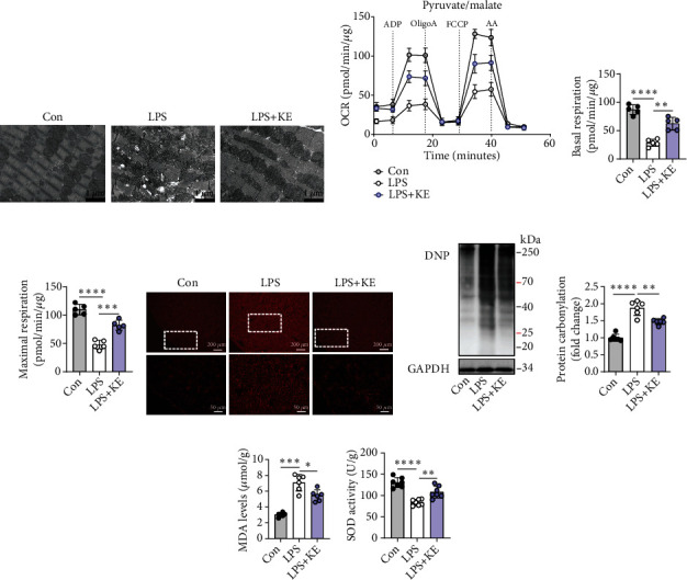
Figure 2.

Ketone bodies mitigated cardiac mitochondrial dysfunction and oxidative stress in septic cardiomyopathy. (a) Representative transmission electron microscopy images of the Con, LPS, and LPS + KE heart sections (n = 5 per group). Scale bar = 1 μm. (b–d) The oxygen consumption rate (OCR) curve in the presence of pyruvate and malate of the isolated mitochondria from the mouse hearts (b). The ADP-stimulated OCR (c) and FCCP-stimulated OCR (d) (n = 5 per group). (e) Representative images of dihydroethidium (DHE) staining in Con, LPS, and LPS + KE hearts (n = 50 fields, from 3 hearts per group). Scale bar = 200 μm or 50 μm. (f and g) Western blotting of 2,4-dinitrophenylhydrazone (DNP) in the heart tissues from Con, LPS, and LPS + KE mice (f) (n = 5 per group). The protein levels of DNP were normalized to the GAPDH levels, and the data were expressed as the fold change relative to the control (g) (n =5 per group). (h) Malondialdehyde (MDA) levels in the heart tissues (n = 5 per group). (i) Superoxide dismutase (SOD) activity in the heart tissues (n = 5 per group). Con: Control; LPS: lipopolysaccharide; KE: ketone ester; ADP: adenosine diphosphate; Oligo: oligomycin A; FCCP: trifluorocarbonyl cyanide phenylhydrazone; and AA: antimycin A. Data are presented as the mean ± SD. Statistical comparisons were conducted using one-way ANOVA, followed by Tukey's multiple comparisons test (c, d, g, h, and i). The exact P values were reported for the indicated comparisons, with P < 0.05 considered statistical significance. ∗P < 0.05, ∗∗P < 0.01, ∗∗∗P < 0.001, and ∗∗∗∗P < 0.0001 for the indicated comparisons.
