
Figure 3.
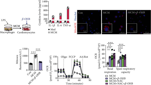
β-OHB alleviated oxidative stress and enhanced aerobic respiration in H9C2 cells exposed to MCM. (a) Schematic representation of the experimental protocol. (b) The levels of inflammatory cytokines in LPS-stimulated macrophage conditioned medium (MCM) (n = 5) and standard cell culture medium (DMEM, Med) with 10% fetal calf serum. (c). Representative fluorescent images of H9C2 cells stained with MitoSOX red after the indicated treatments. Scale bar = 100 μm. The nuclei were visualized with Hoechst staining. Scale bar = 10 μm. (d). The fluorescence intensity of MitoSOX-stained H9C2 cells after the indicated treatments (n = 6 per group). (e and f) The oxygen consumption rate (OCR) curve in the presence of pyruvate and L-glutamine in H9C2 cells after the indicated treatments (e) and the basal respiration and spare respiratory capacity (OCRFCCP–OCRbasal) (f) (n = 5–6 per group). Oligo: Oligomycin A; A A./Rot: antimycin A and rotenone. Data are presented as the mean ± SD. Statistical comparisons were conducted by one-way ANOVA, followed by Tukey's multiple comparisons test (b, d, and f). The exact P values are reported for the indicated comparisons, and P < 0.05 indicates statistical significance. ∗∗P < 0.01 and ∗∗∗P < 0.001 for the indicated comparisons.