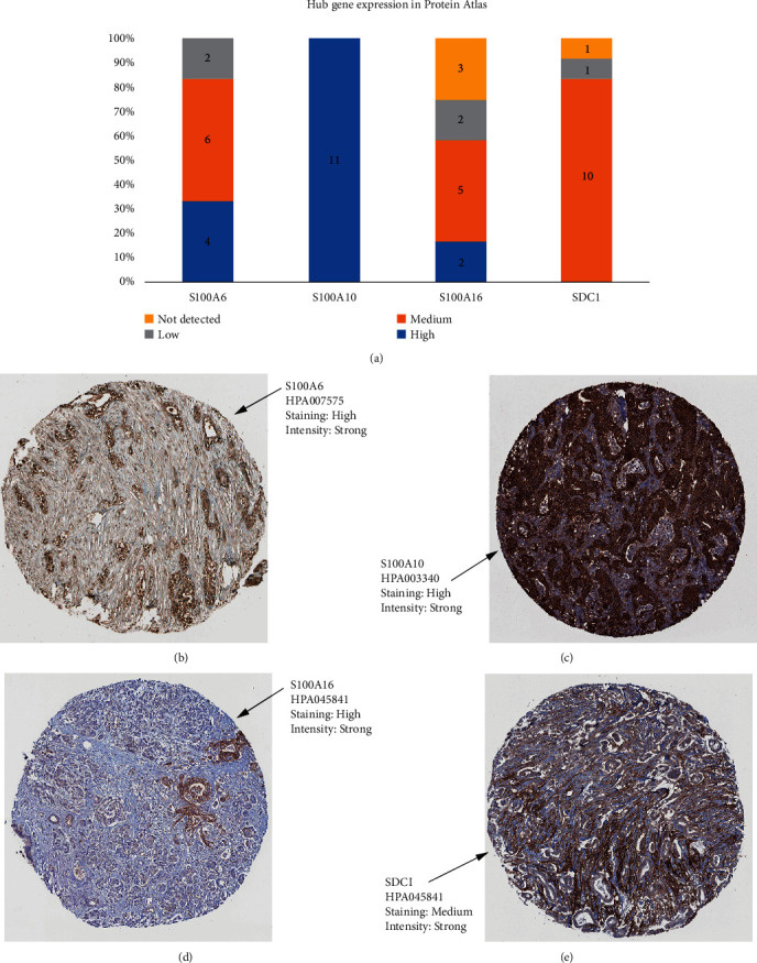
Figure 7.

Detection of hub gene expression by immunochemistry in human pancreatic cancer human tissue. (a) Histogram of mucin expression in PDAC samples from Protein Atlas (http://www.proteinatlas.org/). In total, 11–12 samples were analyzed for S100A6, S100A10, S100A16, and SDC1. Immunohistochemistry (IHC) staining was evaluated as high/medium/low staining or not detected. (b) Representative IHC stainings for S100A6. (c) Representative IHC stainings for S100A10. (d) Representative IHC stainings for S100A16. (e) Representative IHC stainings for SDC1. All hub genes showed a membrane and/or cytoplasmic staining in tumor cells.
