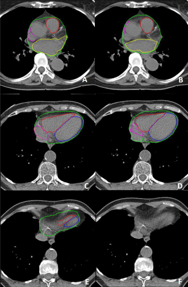
Fig. 4.

Example of poor segmentations in the cranial (A & B) and caudal (C - F) part of the heart with on the left the ground truth and on the right the predicted contours by the deep learning pipeline. Whole heart(WH) (green), left ventricle (LV) (blue), right ventricle (RV) (red), left atrium (LA) (yellow), right atrium (RA) (purple). Note the largely missed right atrium (B & D) and the overestimation of the left atrium (B). Image F shows the most caudal slice being misclassified as containing no cardiac structures
