Figure 1.
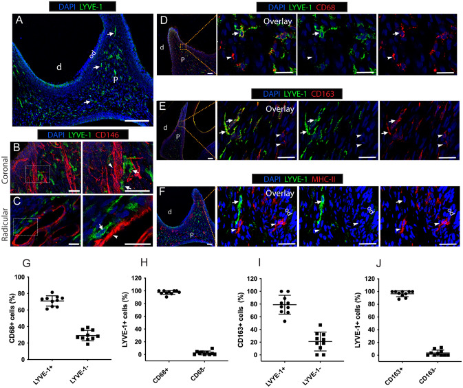
The phenotype of resident LYVE-1+ macrophages in dental pulp tissue of rats (8 weeks old). (A) Immunofluorescence staining of LYVE-1 in rat molar sections. Arrows: LYVE-1+ macrophages. (B,C). Double immunofluorescence staining of LYVE-1 (green) and endothelial cells marker CD146 (red) in rat molar sections. Arrows: LYVE-1+ macrophages; arrowheads: blood vessels. (D) Double immunofluorescence staining of LYVE-1 (green) and pan-macrophage marker CD68 (red) in rat molar sections. Arrows: LYVE-1+CD68+ macrophages; arrowheads: LYVE-1-CD68+ macrophages. (E) Double immunofluorescence staining of LYVE-1 (green) and M2 macrophage marker CD163 (red) in rat molar sections. Arrows: LYVE-1+CD163+; arrowheads: LYVE-1−CD163+ macrophages. (F) Double immunofluorescence stained of LYVE-1 (green) and antigen-presenting cell marker MHC-II (red) in rat molar sections. Arrows: LYVE-1+MHC-II− macrophages; arrowheads: LYVE-1-MHC-II+ macrophages. (G–J). Percentages of macrophage subsets by cell counting (mean ± SD; n = 10). Images are representative of at least 3–4 different samples for each examined condition. d dentin, od odontoblast layer, p dental pulp. Scale bar 200 µm (A) and 50 µm (B–F).

