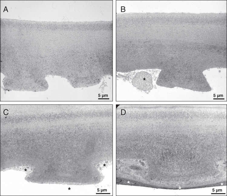
Fig. 1. Transmission electron microscopy images of corneal tissue specimens.
Descemet’s membrane of Patient 2 shows typical ultrastructural features of cornea guttata (A). No overt differences can be observed when comparing with the morphology of Descemet’s membrane removed from the fellow eye (B). Descemet’s membrane of Patient 3 also shows typical guttae. No differences can be discerned between the treated central (C) and the untreated peripheral cornea (D). Asterisks show corneal endothelial cells.

