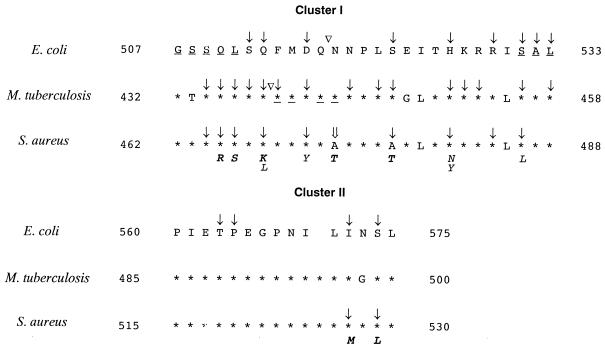
FIG. 2.
Alignment of E. coli, Mycobacterium tuberculosis, and S. aureus rpoB sequences representing clusters I and II (1–3, 11, 14, 25, 26). The amino acid alignment is presented in a single-letter code. Asterisks symbolize identity to E. coli sequence. Positions involved in rifampin resistance are marked; mutations are indicated by downward-pointing arrows; insertions are indicated by upside-down triangles; and deletions are underlined. The new Rifr mutation site found in this study is indicated by a double downward-pointing arrow. Amino acid substitutions involved in Rifr found in this study are presented in italics. Amino acid substitutions not previously described are indicated in boldface.

