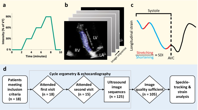
Fig. 1.
Study overview. a Heart failure patients were subjected to echocardiography during exercise at various intensity levels of the ventilatory threshold (VT). b, c This was followed by segmentation and and automated analyses of septal systolic rebound stretch (SRSsept). d Flowchart of ultrasound data processing; from cycle ergometry to speckle tracking-based strain analysis. Legend: AVC aortic valve closure, LA left atrium, LV left ventricle, RV right ventricle, SDI septal discoordination index, SRSsept septal rebound stretch of the septum, VT ventilatory threshold

