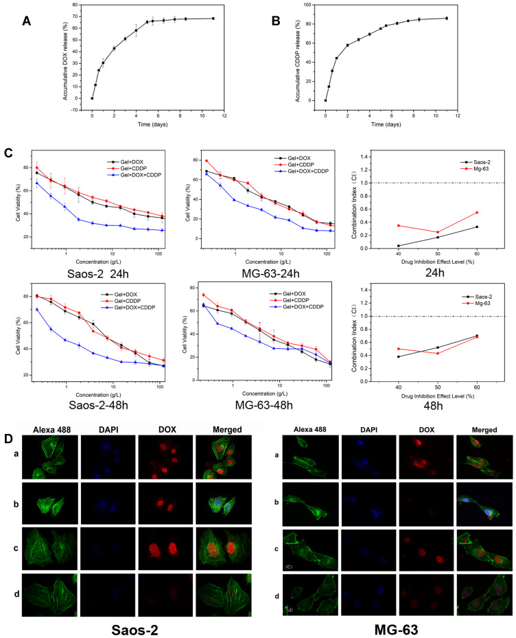
Figure 2.
(A) DOX and (B) CDDP release profiles from the PLGA-PEG-PLGA hydrogels with the drug concentration of 1 mg/mL in PBS at 37 °C in vitro. (C) In vitro cytotoxicities efficacy of DOX and CDDP loaded hydrogels against Saos-2 and MG-63 cells and combination index of DOX and CDDP co-loaded hydrogels after incubation for 24h and 48h. (D) Cellular DOX uptake: ClSM microimages of Saos-2 and MG-63 after incubated with a) DOX, b) Gel+DOX, c) DOX+CDDP, d) Gel+DOX+ CDDP. The cellular cytoskeleton and nuclei of cells were stained with Alexa 488 (green) and DAPI (blue) respectively.

