
Figure 2: Ipsilateral and contralateral projections from the superior colliculus (SC) define two zones of the mouse pulvinar nucleus.
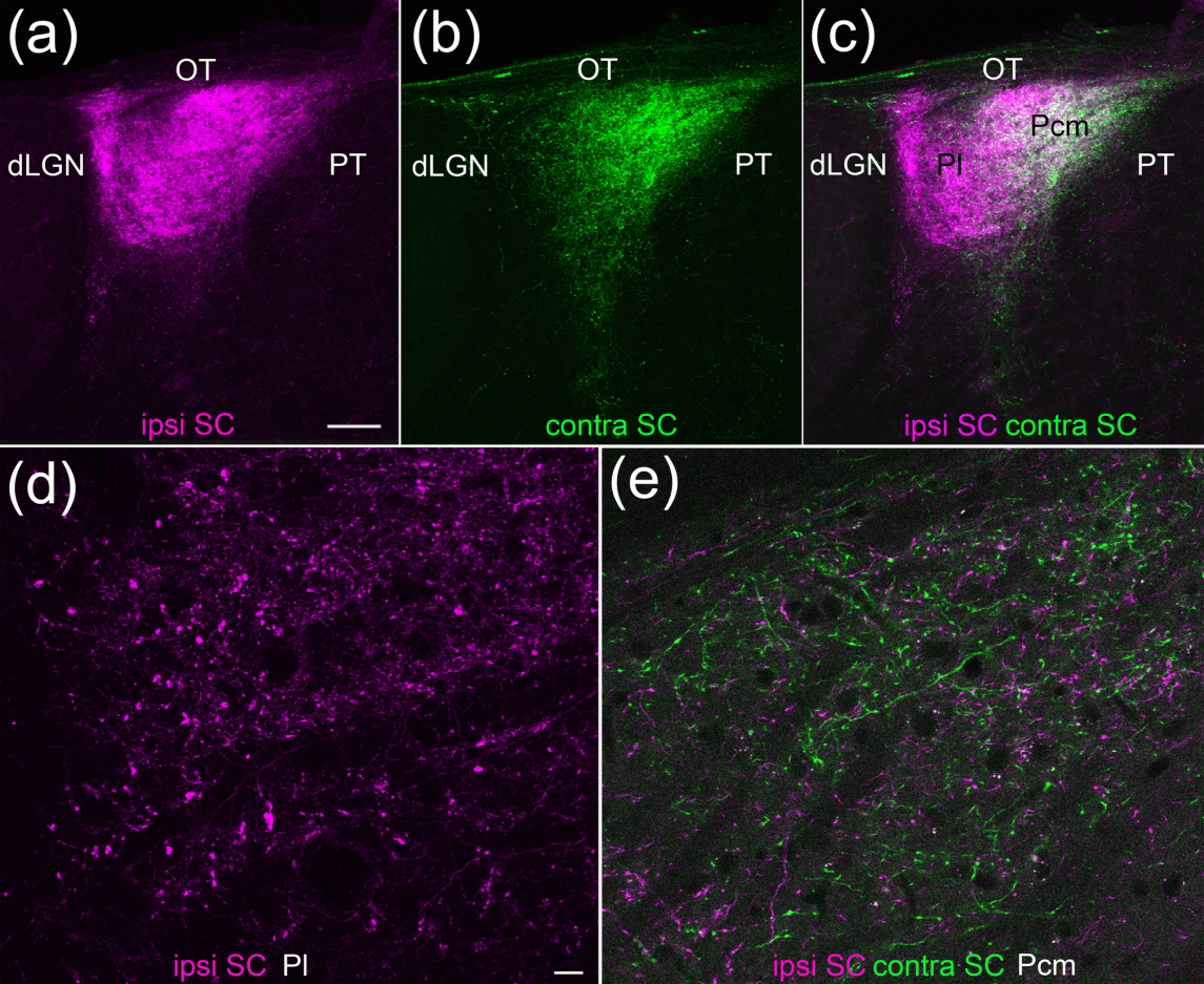
Two different virus injections were placed in the left and right SC and confocal images of coronal sections of the pulvinar nucleus were collected. (a-c) The lateral pulvinar (Pl) is primarily innervated by ipsilateral SC terminals (a,c magenta), while the caudal medial pulvinar (Pcm) is innervated by both ipsilateral and contralateral (b,c green) SC terminals (5 μm optical images, 60 μm stack). Scale bar = 100 μm. d,e) High magnification images of virus-labeled tectopulvinar terminals in the Pl (d) and Pcm (e). 1 μm optical images, 6 μm stacks. Scale bar = 10 μm and applies to d and e. dLGN, dorsal lateral geniculate nucleus, OT, optic tract, PT, pretectum.