
Figure 1.
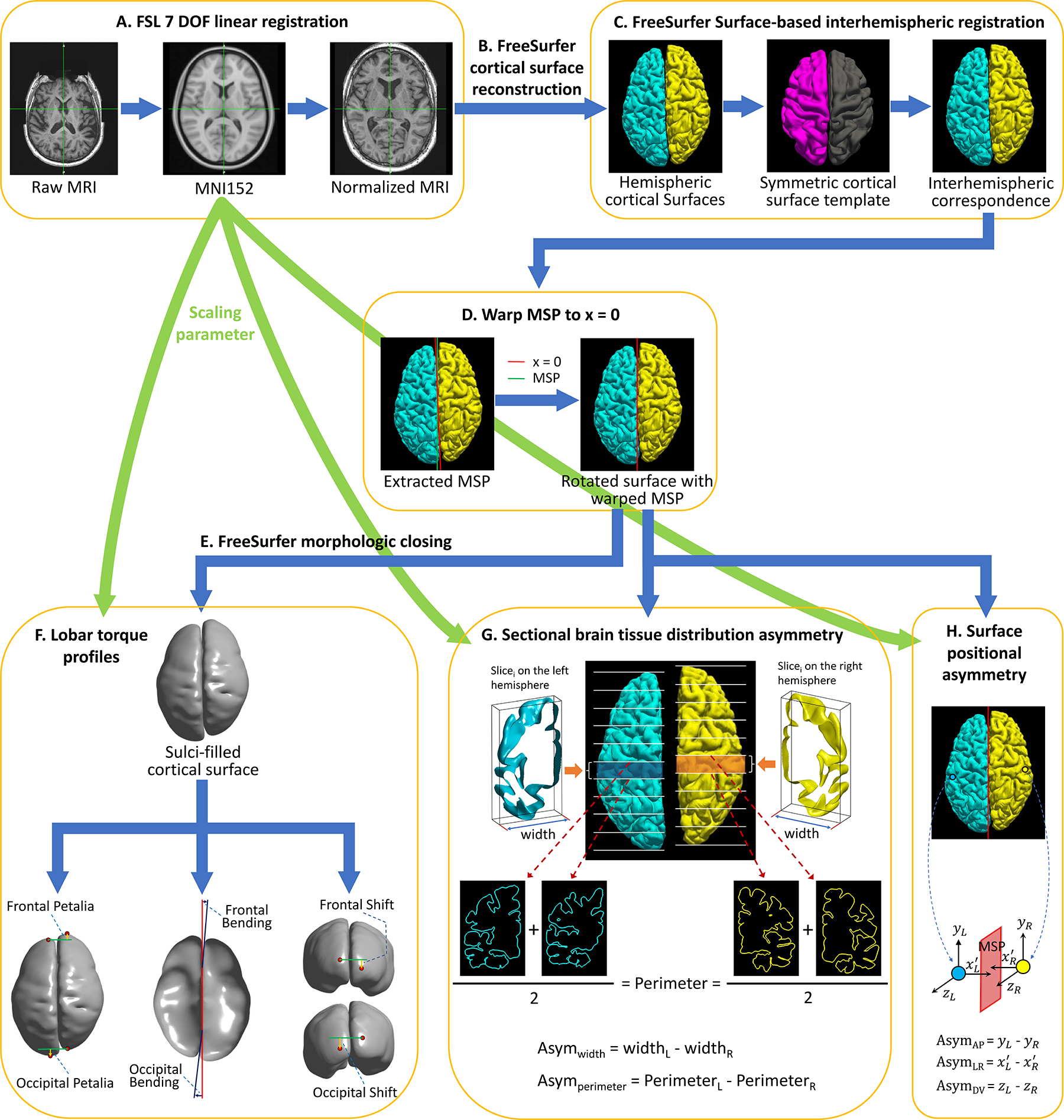
Workflow for computing complex brain torque profiles. A: FSL preprocessing for normalizing the native brain volume to the standard MNI152 template using a 7 degrees of freedom (DOF) transformation. B: FreeSurfer processing on the spatially normalized brain volume to reconstruct cortical hemispheric surfaces. C: FreeSurfer surface-based interhemispheric registration to establish vertex-wise interhemispheric correspondence. D: Correcting for mis-registration of the brain mid-sagittal plane (MSP) by rotating the brain surface with an 3D angle between MSP and the plane x = 0. MSP was extracted as the plane best fitting the two hemispheric medial surfaces. E: FreeSurfer morphologic closing to fill sulci and smooth the cortical surface. F: Computing petalia and shift as the respective displacements of the left and right frontal/occipital extreme points along the antero-posterior and dorso-ventral axes respectively. Estimating bending as the angles between the planes best fitting the interhemispheric fissure in the frontal/occipital regions and MSP. G: Measuring left-right asymmetries in hemispheric width (Asymwidth) and perimeter (Asymperimeter) in 60 contiguous coronal brain slices. Sectional hemispheric width and perimeter was measured as the bounding box width and the average pial surface length at two ends of each brain slice respectively. H: Computing interhemispheric surface positional asymmetries along the left-right axis (AsymLR) as vertex-wise differences in the distances to MSP on the x-axis, and asymmetries along the antero-posterior (AsymAP) and dorsal-ventral axis (AsymDV) as the vertex-wise relative displacements on the y- and z-axes respectively.