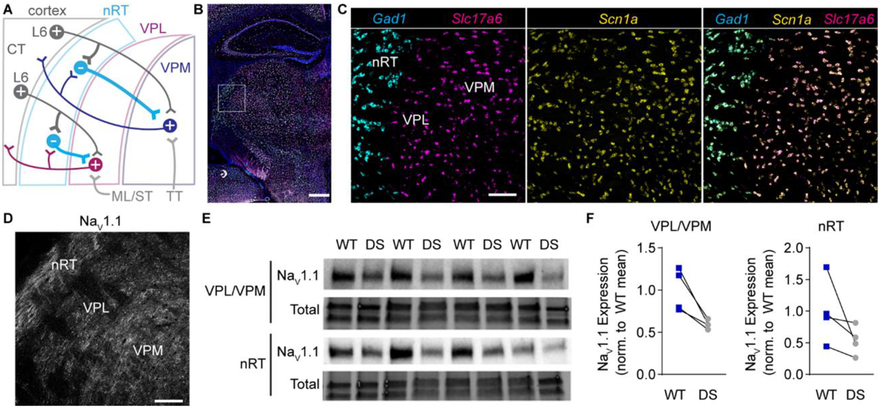
Figure 1. Scn1a mRNA and NaV1.1 protein expression in the somatosensory thalamus.

A. A circuit diagram illustrates somatosensory corticothalamic (CT) circuit connectivity. Layer 6 (L6) glutamatergic CT neurons innervate nRT, VPL, and VPM neurons. GABAergic nRT neurons innervate VPL and VPM neurons, which send glutamatergic projections to the cortex and collaterals to the nRT. Ascending glutamatergic sensory afferents from the medial lemniscus and spinothalamic tract (ML/ST) innervate VPL neurons and the trigeminothalamic tract (TT) innervates VPM neurons. B. A representative 20X tiled image of a coronal mouse brain section shows Scn1a (yellow), Gad1 (cyan), and Slc17a6 (magenta) mRNA labeled by FISH with DAPI counterstain (blue). Scale bar: 500 μm. C. 20X images of the boxed region in panel B show that Gad1+ and Slc17a6+ neurons express Scn1a mRNA. D. A representative 20X image shows NaV1.1 immunolabeling in the nRT, VPL, and VPM. Scale bar: 100 μm (C, D). E. A western blot shows NaV1.1 and total protein expression in nRT and VPL/VPM tissue punches from WT and DS mice (n = 4 littermate pairs). F. NaV1.1 protein levels were quantified by densitometry and normalized
