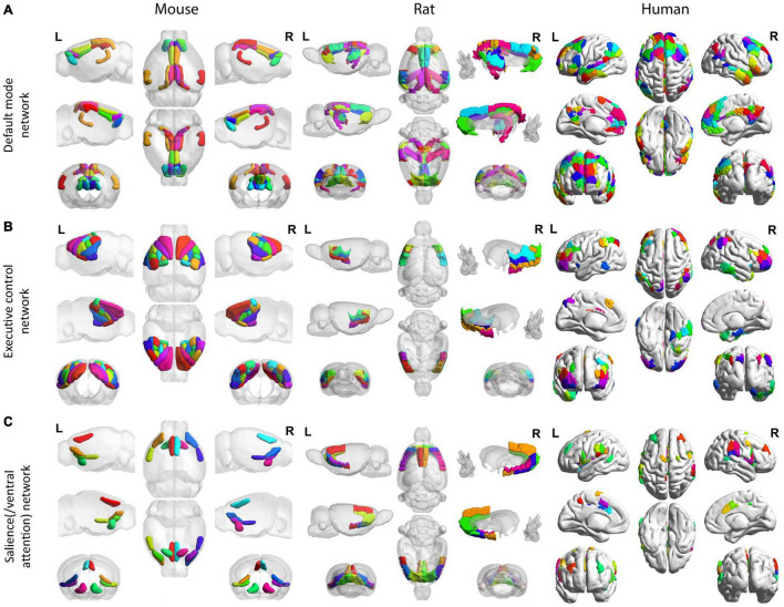
FIGURE 3.
Homologous default mode, executive control, and salience (/ventral attention) networks across mice, rats, and humans. All mouse (left), rat (middle), and human (right) networks are demonstrated on the Allen Institute for Brain Science mouse atlas (Lein et al., 2007), the SIGMA anatomical atlas (Barrière et al., 2019), and the Schaefer-Yeo atlas (Schaefer et al., 2018), respectively. For the DMN (A), the mouse (left) follows the region coverage exported in Grandjean et al. (2020); the rat (middle) follows the region coverage exported in Hsu et al. (2016); the human brain (right) shows the default network in Yeo et al. (2011). For the executive control network (B), the mouse follows the region coverage of lateral cortical network exported in Grandjean et al. (2020); the rat (middle) follows the lateral cortical network coverage exported in Schwarz et al. (2013) except for the anterior secondary motor cortex, which cannot be isolated from the SIGMA anatomical atlas (Barrière et al., 2019); the human brain (right) shows the frontoparietal network in Yeo et al. (2011) and Schaefer et al. (2018). For the salience(/ventral attention) network (C), the mouse (left) follows the salience network commonly found in Sforazzini et al. (2014) and Mandino et al. (2021); the rat (middle) follows the salience network coverage converged by functional and anatomical connectivity from the ventral anterior insular division (Tsai et al., 2020); and the human brain (right) shows the salience/ventral attention network reported in Yeo et al. (2011) and Schaefer et al. (2018). All mouse (left), rat (middle), and human (right) networks are demonstrated on the Allen Institute for Brain Science mouse atlas (Lein et al., 2007), the SIGMA anatomical atlas (Barrière et al., 2019), and the Schaefer-Yeo atlas (Schaefer et al., 2018), respectively. All figures were created by BrainNet Viewer (Xia et al., 2013).

