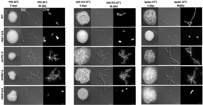
Figure 1.
Colony and cellular morphology of SAGA mutants in C. albicans. 1:10 serial dilution of overnight culture of mutants were spotted on to yeast-growing conditions—YPD agar and hyphae-inducing conditions—10% fetal calf serum media (FCS) and Spider media plates. The colony morphology was assessed after 5 days of incubation. Cells from liquid media were inoculated at starting OD600 of 0.2 and grown in liquid YPD, 10% FCS supplemented YPD and Spider medium, at 220 rpm, and 30°C or 37°C for 4 h for normal growing strains (WT, ngg1Δ/Δ and ubp8Δ/Δ) and 6 h for slow growing strains (spt7Δ/Δ and spt8Δ/Δ). The cells were washed with 1× PBS twice and stained with 2 µg/ml calcofluor white (CFW). The cells were observed with the Leica DM6000 microscope at ×100 magnification-DIC (Differential Interference Contrast). Scale bar = 15 µm. spt7Δ/Δ and spt8Δ/Δ appear more hyphal compared to control and are mostly in pseudo-hyphal state in inducing and non- inducing media whereas ngg1Δ/Δ and ubp8Δ/Δ appear in yeast locked state. The control switches its morphology upon changed conditions while the SAGA mutants remain in their initial states upon induction.

