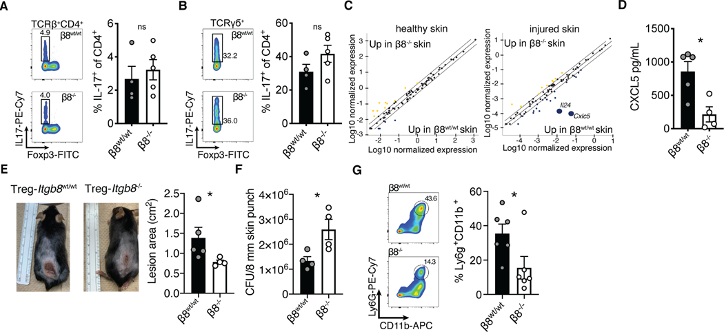
Fig. 4. αvβ8+ Tregs promoted innate skin inflammation.
(A, B) Tregs from Foxp3Cre-ERT2Itgb8f/f mice treated with either vehicle or tamoxifen were phenotyped by flow cytometry for IL-17 expression in skin CD4+ αβT cells (A), or γσT cells (B) after epithelial injury. (C) Whole skin quantitative PCR array analysis comparing cytokine and chemokine gene expression levels between β8wt/wt and β8−/− healthy, or tape strip injured mice. (D) Whole skin biopsy lysate ELISA for CXCL5. (E, F) Treg β8wt/wt and β8−/− mice intradermally infected with 5×10⁷ CFU Staphylococcus aureus (USA300) were monitored for six days before an 8 mm punch biopsies surrounding lesions were excised for bacterial colony forming unit analysis. (G) Representative flow plots and quantification of neutrophil accumulation in skin following Staphylococcus aureus infection. Data are representative of at least three independent experiments, n = 4–7 mice per group; error bars are SEM. Statistical analysis was performed with a unpaired Student’s two tailed t test. *P<0.05.

