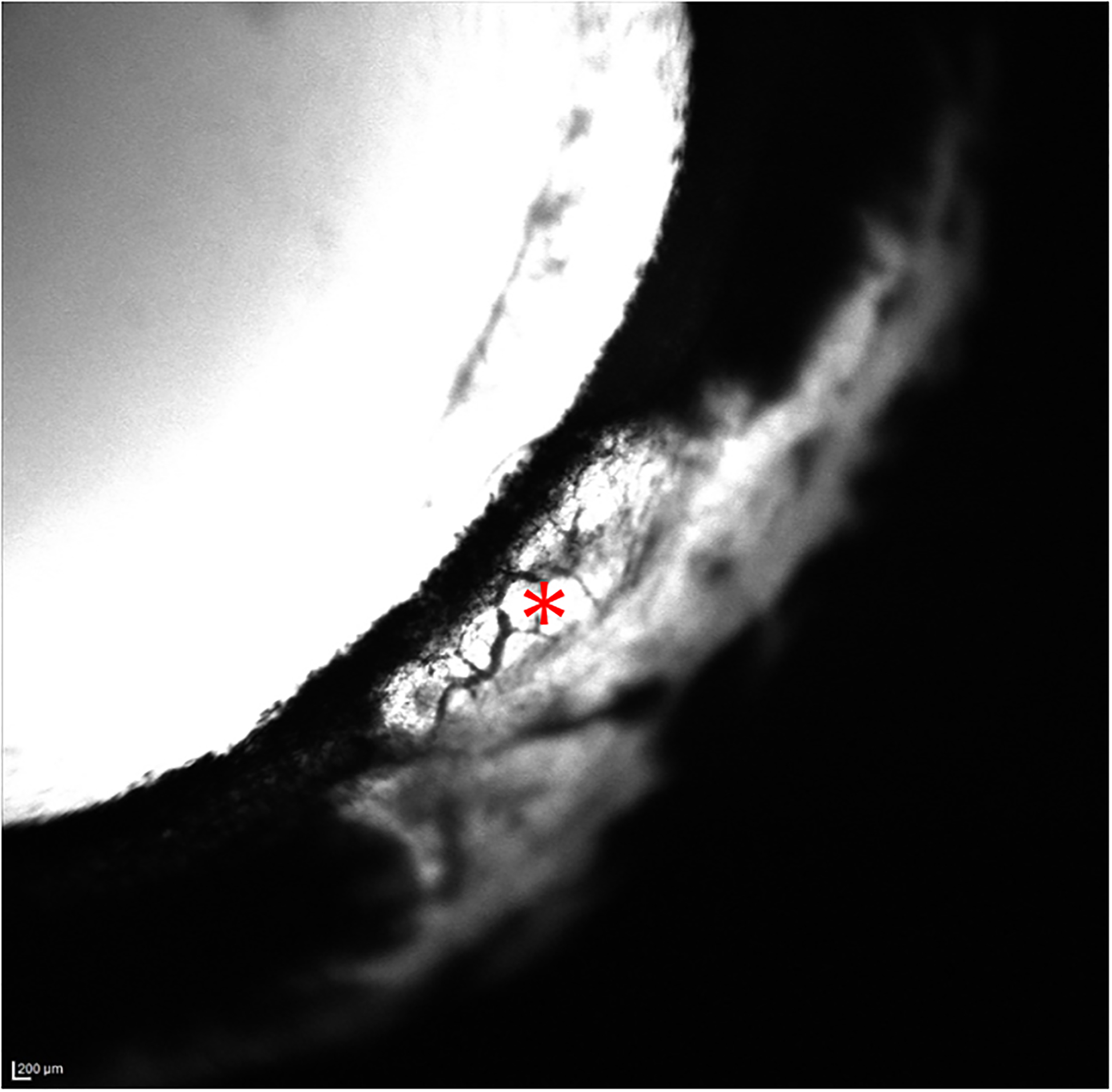
Figure 3.

Aqueous Angiography (AA) images of the temporal quadrant of the left eye (OS) acquired with indocyanine green (ICG) for glaucomatous eye G2. There are no areas of well-defined angiographic signal several minutes post-infusion of ICG. The white area of sclera appeared to represent fluorescence of dye in aqueous humor that was visible through thin sclera in this region, small perilimbal vessels that do not contain fluorescent tracer are visualized in these images as dark, branching structures against a fluorescent background (*), even when sensitivity settings were increased.
