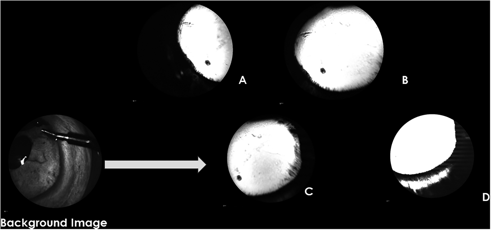
Figure 4.

Aqueous Angiography (AA) images obtained glaucomatous eye G4 following infusion of fluorescein. The background image shows the orientation of the right eye with the anterior chamber maintainer in the dorsonasal quadrant. A-D show images of different quadrants acquired around the 10-minute time point following tracer infusion (A=temporal, B=nasal, C=nasal, D=ventral). There are no areas of well-defined AA signal in any quadrant. The “white”, hyperfluorescent area in D reflects scleral diffusion of fluorescein but is not considered representative of an AA signal.
