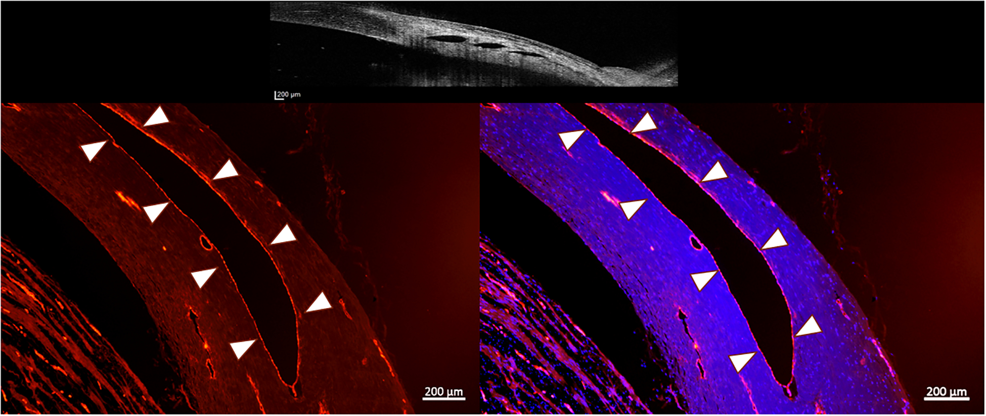
Figure 7.

Fluorescence photomicrographs of perilimbal sclera from a representative normal canine eye. The red immunofluorescent labeling by von Willebrand factor of vascular endothelium on the inner surface of each lumen (outlined by white arrow heads) confirms the vascular nature of these lumens (positive control). Right image, merged with blue fluorescent DAPI-stained nuclei (lumen highlighted with white arrow heads). Top image is an OCT image showing the scleral lumens being identified with positive controls. (Images intentionally overexposed to facilitate visualization of tissue morphology). (Bar Markers 200μm, 5X magnification)
