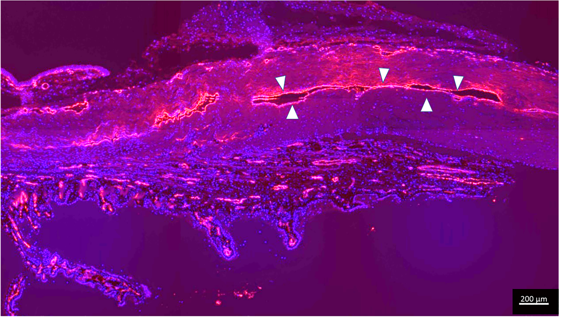
Figure 8.

Immunolabeling for endothelial marker confirms presence of collapsed scleral vessels in glaucomatous dog G2 which had no angiographic signal. Endothelial cells are highlighted in red (outlined in white arrow heads), lining the collapsed lumens with DAPI (blue nuclear counterstain). (Bar marker 200μm)
