Figure 9.

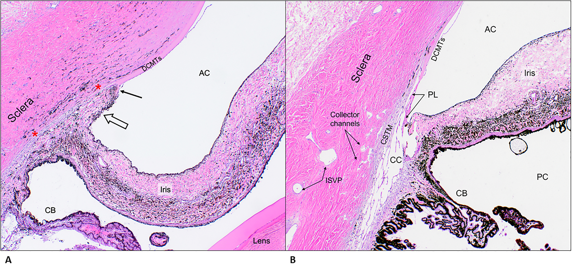
A. Representative photomicrograph of a section of the iridocorneal angle from patient G5, a Leonberger, illustrating features consistent with primary glaucoma. Note an iris-like tissue (open arrow) crossing over the iridocorneal angle opening contacting the arborizing (fibrillating) end of Descemet’s membrane (black arrow) replacing the normal pectinate ligaments and ciliary cleft opening. Also, in the region between the red asterixis, note the absence of an identifiable ciliary cleft and trabecular meshwork tissue and paucity of angular aqueous plexus structures. H&E, 4X objective. B. Representative photomicrograph of a section of the iridocorneal angle from a control dog illustrating the normal anatomical features. H&E, 4X objective. AC= anterior chamber; DCMTs= Descemet’s membrane; PL= pectinate ligaments; CSTM= corneo-scleral trabecular meshwork; CC= ciliary cleft; CB= ciliary body; PC= posterior chamber; ISVP= intra-scleral venous plexus
