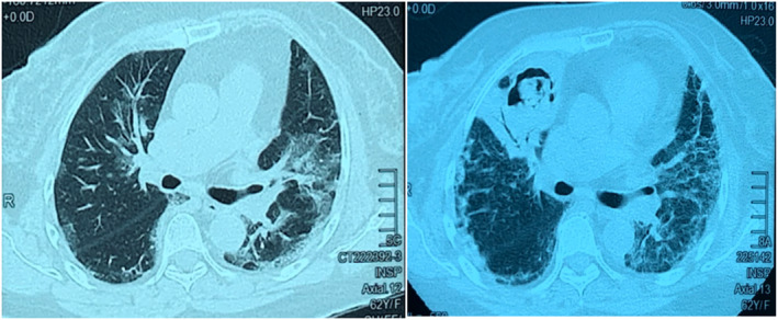
FIGURE 1.

Patient 1. Computed tomography (CT) thorax on day 8 of COVID‐19 infection (left panel) showed bilateral peri‐bronchovascular ground‐glass opacity. On day 40 of COVID‐19 infection (right panel), CT showed new cavitating lesion at the anterior segment of the right upper lobe with soft tissue within it. Consolidative changes were noted adjacent to the cavity
