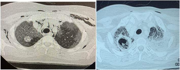
FIGURE 2.

Patient 2. Computed tomography thorax on day 13 of COVID‐19 infection showed peripheral ground‐glass opacity with extensive pneumomediastinum and subcutaneous emphysema. Round heterogenous hyperdensity of the right upper lobe was noted raising the suspicion of infection (tuberculosis, fungal or bacterial) or malignancy, although less likely. On day 32 of COVID‐19 infection, a new cavitating lesion was noted at the right upper lobe where the nodule was seen. Previously seen ground‐glass opacity at the left lung appeared more dense
