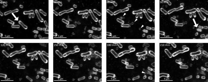
Fig. 1.
Time-lapse microscopy of Paracoccus denitrificans Pd1222 WT treated with 50 ng mL–1 MMC.
White arrows indicate lysed cells. White triangles indicate membrane vesicles (MVs) that were produced as a result of cell lysis. The numbers in the upper left corner indicate the elapsed time (min) when the time point at the beginning of imaging was 0. White: FM4-64 (membrane); Scale bar: 5 μm.

