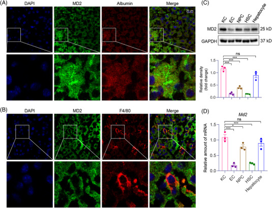
FIGURE 3.

MD2 protein localizes to hepatocytes and hepatic macrophages in liver tissues. (A) Representative immunofluorescence staining images of normal mouse liver tissues showing immunoreactivity to MD2 (green) and hepatocyte‐marker albumin (red). Tissues were counterstained with DAPI (blue). Arrows indicating co‐localization (scale bars = 25 μm). (B) Representative staining images showing immunoreactivity to MD2 (green) and macrophage marker F4/80 used to detect hepatic macrophages (red). Tissues were counterstained with DAPI (blue). Arrows indicating co‐localization (scale bars = 25 μm). (C and D) Primary Kupffer cells (KC), endothelial cells (ECs), non‐parenchymal cells (NPC), hepatic stellate cells (HSCs) and hepatocytes were isolated from mouse livers. The protein (C) and mRNA (D) levels of MD2 in these cells were examined by western blot and qPCR assay, respectively (n = 3; mean ± SEM; ns, not significant; *p < .05, **p < .01 and ***p < .001)
