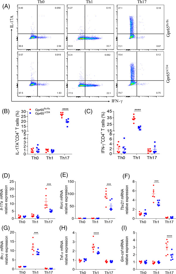
FIGURE 3.

Gpr65ΔCD4CD4+ T cells compromise Th1 and Th17 cell differentiation in vitro. Splenic naïve CD4+ T cells were separated from Gpr65flx/flx and Gpr65ΔCD4 mice (n = 6/group) using anti‐mouse CD4 magnetic beads, and cultured under different polarising conditions. (A) These polarising CD4+ T cells were harvested on day 5, and protein expression of IFN‐γ and IL‐17A was evaluated by flow cytometry. (B and C) Percentages of IFN‐γ +CD4+ and IL‐17A+CD4+ T cells were exhibited in the chart. (D to I) The mRNA levels of Il‐17a, Rorc, Ifn‐γ, Tbx21, Tnf‐α, and Gm‐csf in these different polarising CD4+ T cells were analysed by qRT‐PCR. Data were representative of three independent experiments. Data were presented as mean ± SEM. Statistical analysis was performed with unpaired Student's t tests. ***p < 0.001, ****p < 0.0001
