. 2022 Mar 25;15(1):1–7. doi: 10.3400/avd.ra.21-00055

Fig. 2 (A) Computed tomography angiography showing a stenosed superior mesenteric artery with distal perfusion. There is no embolus nor atherosclerotic changes surrounding the lesion. (B) Nonselective angiography showing isolated superior mesenteric artery dissection (ISMAD). (C) Selective angiography showing ISMAD.
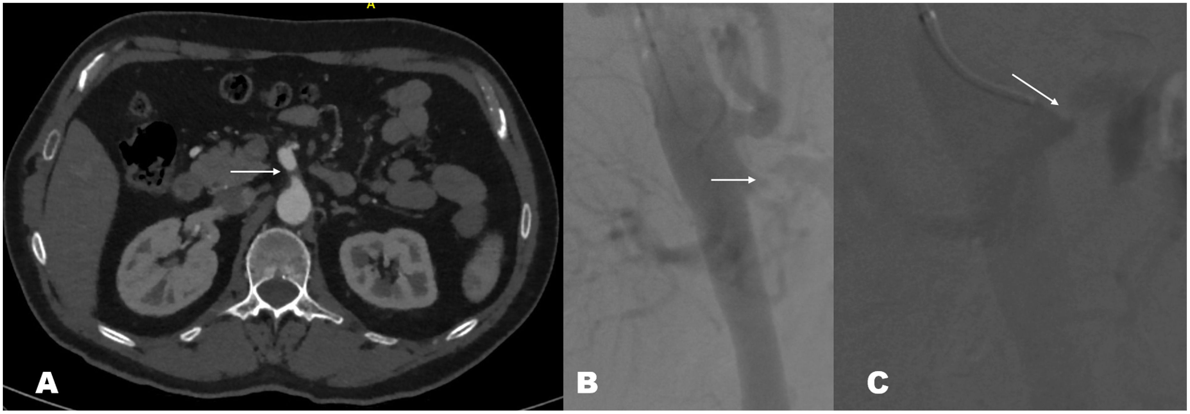
Fig. 2 (A) Computed tomography angiography showing a stenosed superior mesenteric artery with distal perfusion. There is no embolus nor atherosclerotic changes surrounding the lesion. (B) Nonselective angiography showing isolated superior mesenteric artery dissection (ISMAD). (C) Selective angiography showing ISMAD.