


Figure 2: RGN promotes dormancy in vivo.
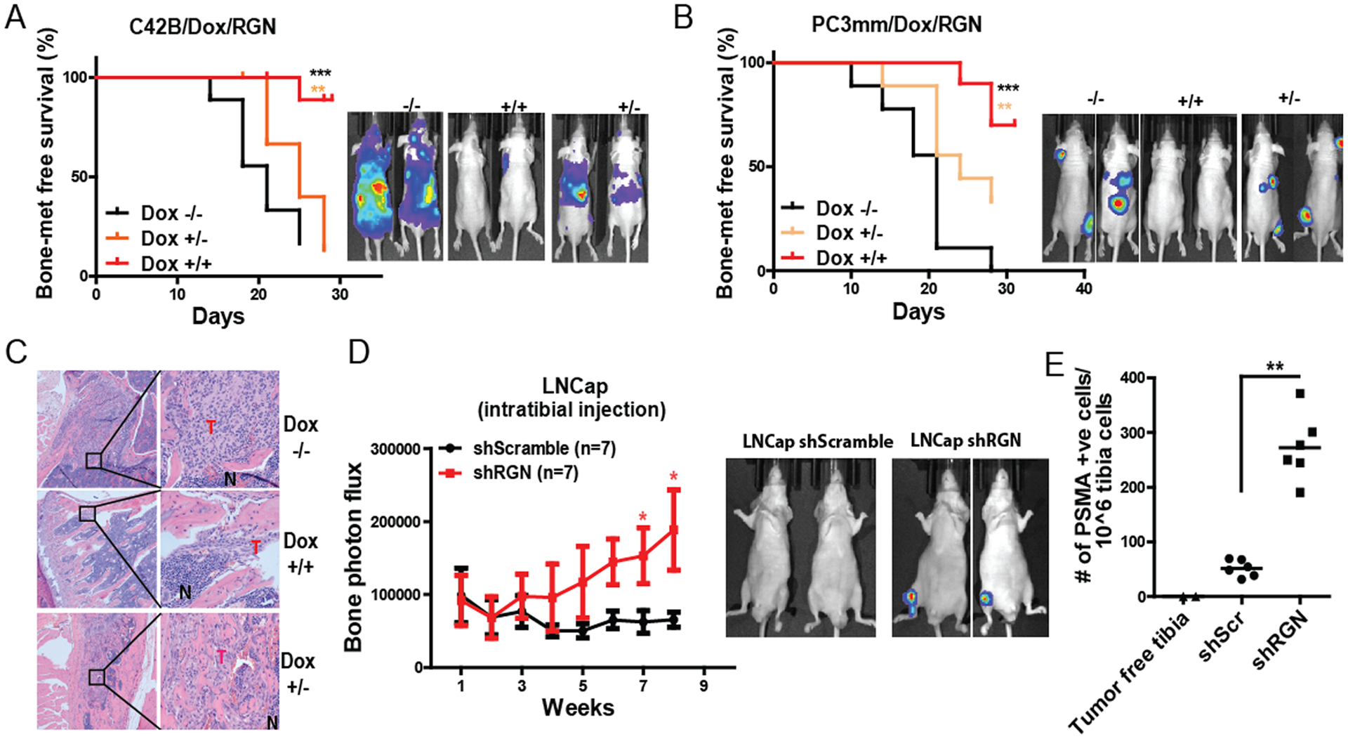
(A) C42B cells were modified to express RGN in doxycycline dependent manner. These cells were systemically implanted into nude mice by intracardiac injection, and kaplan-meier bone metastasis-free survival curve was plot (5 × 10⁵ cells/mouse, n=9/group). −/− group was fed with 5% sucrose water, +/+ group received doxycycline water (2mg/ml in 5% sucrose) and +/− group was fed with doxycycline water for 14 days followed by feeding mice with 5% sucrose water. Panel on the right shows representative bioluminescence image of mice at endpoint. (B) PC3mm cells were modified to express RGN in a doxycycline dependent manner and transplanted to nude mice via intracardiac injection (5 × 10⁵ cells/mouse, n=9/group). −/− group was fed with 5% sucrose water, +/+ group received doxycycline water (2mg/ml in 5% sucrose) and +/− group was fed with doxycycline water for 12 days followed by feeding mice with 5% sucrose water. Panel on the right shows representative bioluminescence image of mice at endpoint. The bone-metastasis free survival plots in Figure A–B were analyzed by log-rank (Mantel-cox) test. (C) Representative H&E staining of bone sections from mice in panel A. (D) LNCap shScramble and LNCap shRGN cells were injected in nude mice and tumor growth in tibia was examined by IVIS imaging (n=7/group, 150,000 cells/tibia). Representative images of IVIS imaging are shown in the right panel. The data collected at each time point was analyzed using unpaired t-test. (E) Human-specific PSMA antibody staining was performed in the tibia flushed cells from mice in panel E by flow cytometry. Unpaired t-test was used to analyze the data. *, P-value<0.05, ** P-value<0.01 and ***, P-value<0.0001.