Figure 1.
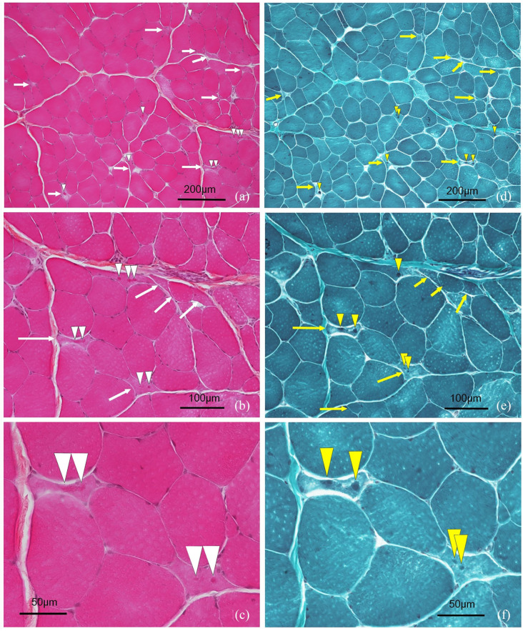
Muscular histopathological changes. Transverse sections of a fresh-frozen muscle biopsy from the left biceps muscle stained with (a–c) haematoxylin, eosin and saffron (HES) and (d–e) Gomori trichrome navy blue staining (GT). (a,d) × 100; (b,e) × 200; (c,f) × 400. Atrophic fibres were observed (yellow and white arrows) and most contained inclusions, which appeared amphophilic on HES (white arrowheads) and blue on GT (yellow arrowheads)

