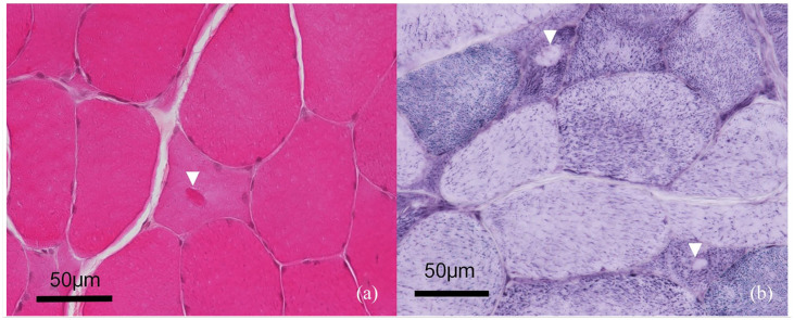
Figure 2.
Morphology of the inclusions. Transverse section of a fresh-frozen muscle biopsy from the left biceps muscle (× 400). (a) Haematoxylin, eosin and saffron staining: a 10 µm intrasarcoplasmic oval shape amphophilic inclusion is located at the centre of a myofibre (arrowhead). (b) Nicotinamide adenine dinucleotide dehydrogenase tetrazolium reductase reaction staining: two atrophic myofibres exhibit similar oval-shaped inclusions in the form of unstained halos (arrowheads)

