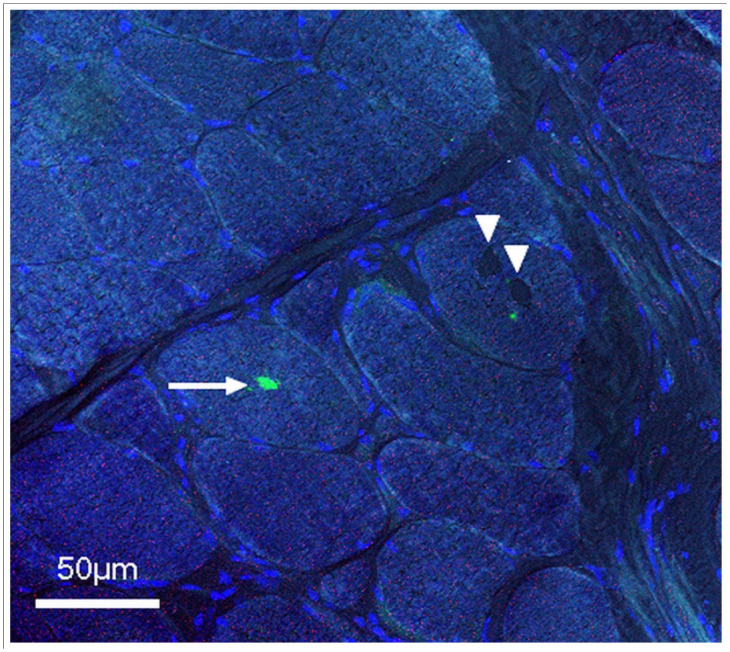
Figure 3.

LC3 immunostainings of a transverse section of a fresh-frozen muscle biopsy from the left biceps muscle (× 400). Inclusions (arrowheads) are distinguishable but are not marked by LC3 antibodies. A macrophage located inside the fibre (arrow) is marked by LC3, suggesting macrophage phagocytosis secondary to fibre necrosis. Nuclei counterstained in blue and phase contrast to depict fibre limits
