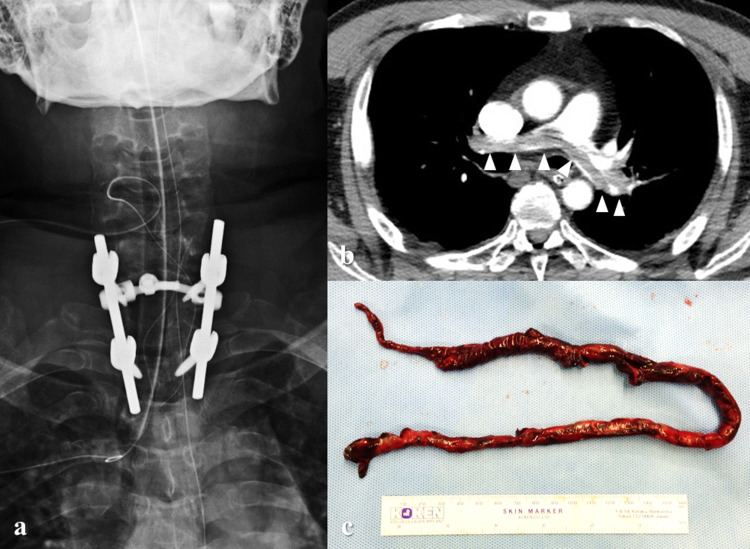
Figure 2. Postoperative images.
(a) X-ray taken immediately after spinal surgery. C7-T2 posterior fusion and C7 and T1 laminectomies were performed. (b) Contrast-enhanced CT at three days after surgery. Extensive defect of contrast medium in the area ranging from the main pulmonary artery to the pulmonary artery bifurcation extending into both trunks (arrowheads). (c) The main pulmonary artery was incised to remove a giant thrombus (39 cm).

