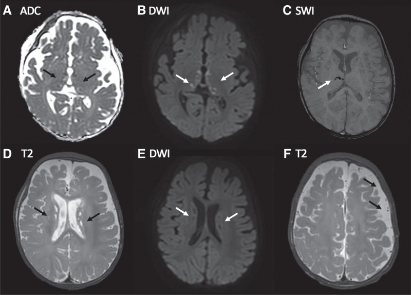
Figure 2.

Presenting features: magnetic resonance imaging (MRI) brain findings. MRI day 2 postadmission: (A) Apparent diffusion coefficient (ADC) map and (B) corresponding diffusion-weighted image (DWI) show multiple punctate lesions in the thalami and globi pallidi that are associated with true diffusion restriction consistent with small acute (likely <7 d old) infarcts (arrows). (C) Susceptibility-weighted image (SWI) showing signal loss in a few of the right thalamic lesions consistent with microhemorrhage (arrow). (D) T2-weighted image demonstrating numerous T2 hyperintense periventricular punctate lesions that have no corresponding restricted diffusion (E) consistent with nonacute infarcts. (F) T2-weighted axial image demonstrating subtle widening of the left frontoparietal subarachnoid space (arrows) likely due to reduced left hemispheric volume consequent upon microscopic remote ischemic white matter/subplate injury postnatally or prenatally.
