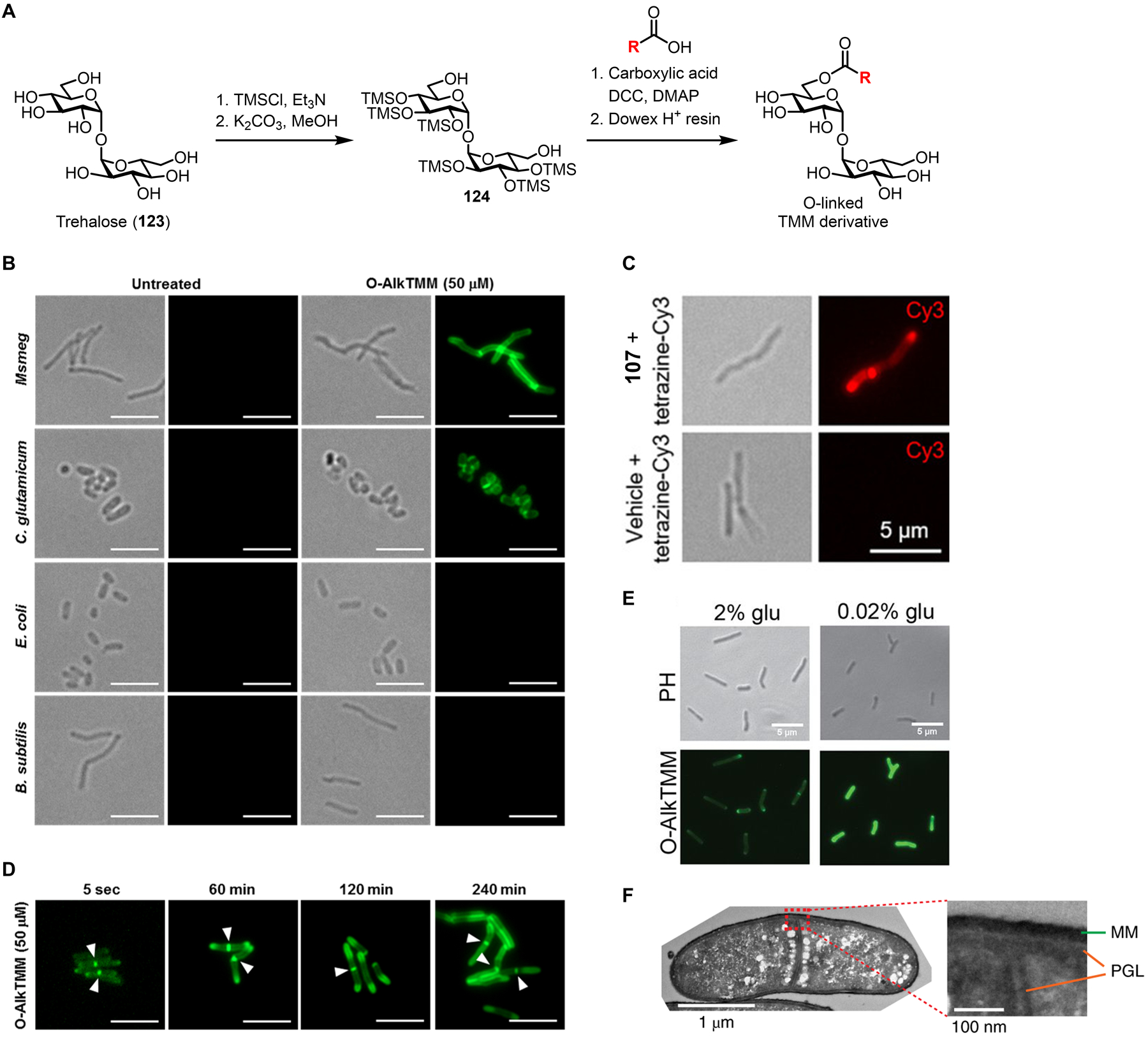
Figure 39.

Labeling and imaging mycolates in mycobacteria using TMM derivatives. (A) General scheme for the synthesis of O-linked TMM derivatives. DCC, N,N’-dicyclohexylcarbodiimide; DMAP, 4-dimethylaminopyridine; TMS, trimethylsilyl. (B) Different species of bacteria were incubated in the presence of 50 μM O-AlkTMM-C7 (113) or left untreated, fixed, reacted with azido-488 via CuAAC, and imaged by fluorescence microscopy. For each condition, left is the transmitted light channel and right is the 488 channel. Scale bars, 5 μm. (C) M. smegmatis was incubated in the presence of 20 μM O-TCO-TMM (117) or left untreated, fixed, reacted with tetrazine-Cy3 for 1 min and imaged by fluorescence microscopy. Left, transmitted light channel; right, Cy3 channel. Scale bars, 5 μm. Reproduced with permission from ref 274. Copyright 2019 Wiley-VCH. (D) M. smegmatis was incubated in the presence of 50 μM O-AlkTMM-C7 (113) for varying durations, fixed, reacted with azido-488 via CuAAC, and imaged by fluorescence microscopy. Arrows indicate sites of septal mycomembrane synthesis in dividing cells. Scale bars, 5 μm. (B) and (D) reproduced with permission from ref 271. Copyright 2016 Wiley-VCH. (E) M. smegmatis was incubated in the presence of 50 μM O-AlkTMM-C7 (113) in 2% (carbon-rich) or 0.02% (carbon-depleted) glucose-supplemented medium, fixed, reacted with azido-488 via CuAAC, and imaged by fluorescence microscopy. Scale bars, 5 μm. Reproduced with permission from ref 309. Copyright 2021 American Society for Microbiology. (F) C. glutamicum was incubated in the presence of 50 μM O-DBF-TMM (120), fixed, photo-oxidized in the presence of diaminobenzidine, stained with osmium tetroxide, and imaged by transmission electron microscopy. The stained mycomembrane (MM) and peptidoglycan layer (PGL) are indicated. Reproduced with permission from ref 275. Copyright 2019 Springer Nature.
