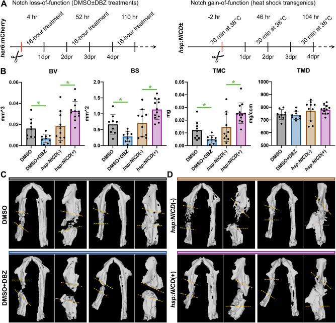
Fig. 2.
Regenerate bone quantity scales with post-operative Notch signaling dose. (A) Complementary approaches were used to assess the role of Notch signaling in mandible regeneration during the initial post-operative period. (B) µCT analyses at 32 dpr demonstrate reduced bone formation following DBZ treatment and enhanced bone formation with hsp:NICD. Notch signaling levels correlate with bone quantity (BV, BS, TMC) but not density (TMD). The four groups represent 9, 9, 10 and 12 animals, from left to right. Each data point represents an individual fish, with bars depicting mean±s.d. *P<0.05 (two-tailed t-tests with Welch's correction). (C,D) Two representative µCT surface renderings are shown for each group to illustrate range, in two orientations offset by 90°: supine, and lateral recumbent with the intact side digitally removed for clarity. Approximate surgical margins are depicted with yellow dashed lines; actual injury margins were carefully delineated slice-by-slice during µCT analysis.

