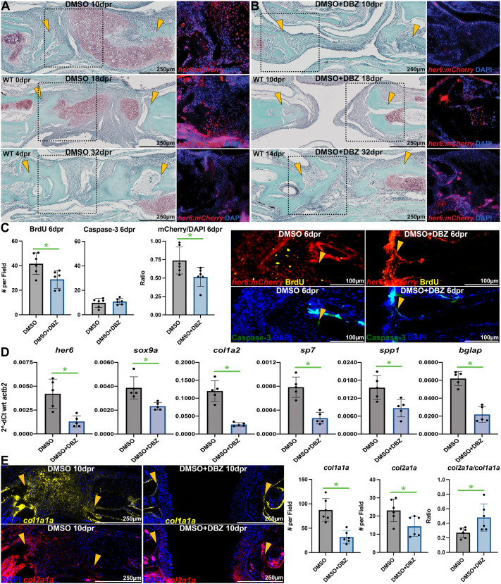
Fig. 3.
Inhibition of Notch signaling represses osteochondral callus formation. (A,B) Safranin O staining (left column) and mCherry IHC (right column) from her6:mCherry reporter zebrafish at 10-32 dpr reveal inhibition of initial callus formation and Notch signaling following three sessions of post-operative DBZ treatment (as in Fig. 2A). Dotted boxes in Safranin O images represent approximate source locations and scale of IHC images from adjacent slides. Yellow arrowheads indicate approximate surgical margins. (C) At 6 dpr, DBZ treatment decreased proliferation (as measured by BrdU staining) of mCherry+ cells at the injury margins, whereas apoptosis (measured by caspase-3 IHC) was not changed (n=6 per group). Representative images are shown on the right. Yellow arrowheads indicate approximate surgical margins. (D) At 10 dpr, 5 days following the conclusion of DBZ treatment, lasting alterations in callus osteochondral gene expression were observed. Each point represents a pool of least 11 animals (n=5 pools in each group). (E) ISH for col1a1a and col2a1a reveals fewer callus cells and increased col2a1a/col1a1a ratio following DBZ treatment (n=6 per group). Each data point represents an individual fish, except for qPCR data, for which each point represents a different pool. Bars depict mean±s.d. *P<0.05 (two-tailed t-tests with Welch's correction).

