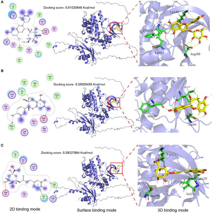
Figure 9.
Molecular docking and protein-ligand interactions. (A) Cyanidin-3- glucoside binding mode with AMPKα; (B) Catechin binding mode with AMPKα; (C) Chlorogenic acid binding mode with AMPKα; The 3D structure of cyanidin 3-O-glucoside, catechin, and chlorogenic acid are shown in yellow, the structure of nearby residues is shown in green, the skeleton of the receptor protein is shown in light blue cartoon, and hydrogen bond is represented by the yellow dotted line.

