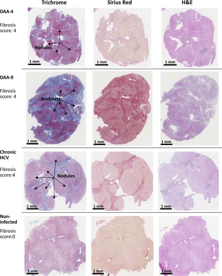
Figure 3.
Examples of trichrome stain, picrosirius red stain, and H&E stain analysis of human liver slices. Arrow indicates the nodule formation in the cirrhosis livers. The Scheuer/Batts-Ludwig method was used for fibrosis scoring, with 0, No fibrosis; 1, portal fibrosis; 2, peri-portal fibrosis; 3, bridging fibrosis; and 4, cirrhosis. The DAA-4 liver tissue was collected 12 months after the completion of 24-week DAA therapy. DAA-9 liver tissue was collected 20 months after the completion of 12-week DAA therapy.

