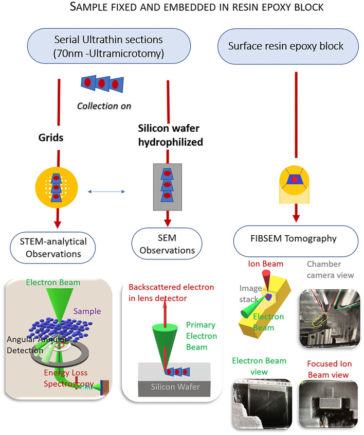
Figure 2.
Methodology workflow of electron microscopy (EM) combined approaches from a unique sample. EM preparation follows a standard protocol. From the same sample, ultrathin sections are collected either on grids for scanning and transmission electron microscopy [(S)TEM] analytical observations or on silicon wafers for scanning electron microscopy (SEM) observations. Tomography focus-ion-beam-SEM (FIB-SEM) is performed on the resin block whose surface was smoothed by the previous ultramicrotomy process. The resulting data gives information on the location and distribution of the endothelial glycocalyx in a volume fraction of the artery.

