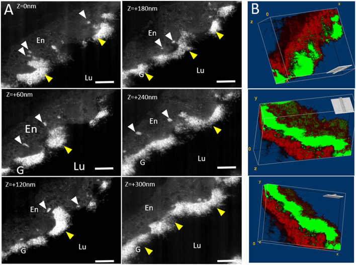
Figure 4.
Tomography 3D FIB-SEM under physiological conditions: (A) Micrographs extracted from FIB-SEM stack at different z-position. Stack images were acquired from 37 “slice and view” images with a step of 10 nm. Glycocalyx (G) (yellow arrows) appears as white dense-packed material at the endothelial (En). White arrows point endothelial vesicles filled with glycocalyx. (B) Volume reconstruction: Glycocalyx is thresholded as green part while the endothelium is in red. Note the presence of glycocalyx inside the intracytoplasmic vesicles. Scale bar = 1 μm.

