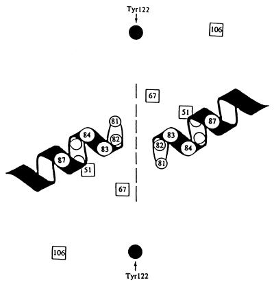
FIG. 7.
Relative positions of resistance alleles in GyrA dimer. The numbers indicate amino acid positions in the E. coli GyrA protein for resistance mutations and for the active center tyrosine (solid circles). The resistance alleles that reside within α-helix 4 are shown as open circles; residues outside the helix that confer resistance are shown as open squares. The dashed line approximates the interface of the two GyrA subunits. DNA is predicted to lay across the protein at an angle from upper left to lower right such that the two helices fit in the major groove of DNA (14).

