FIGURE 5.
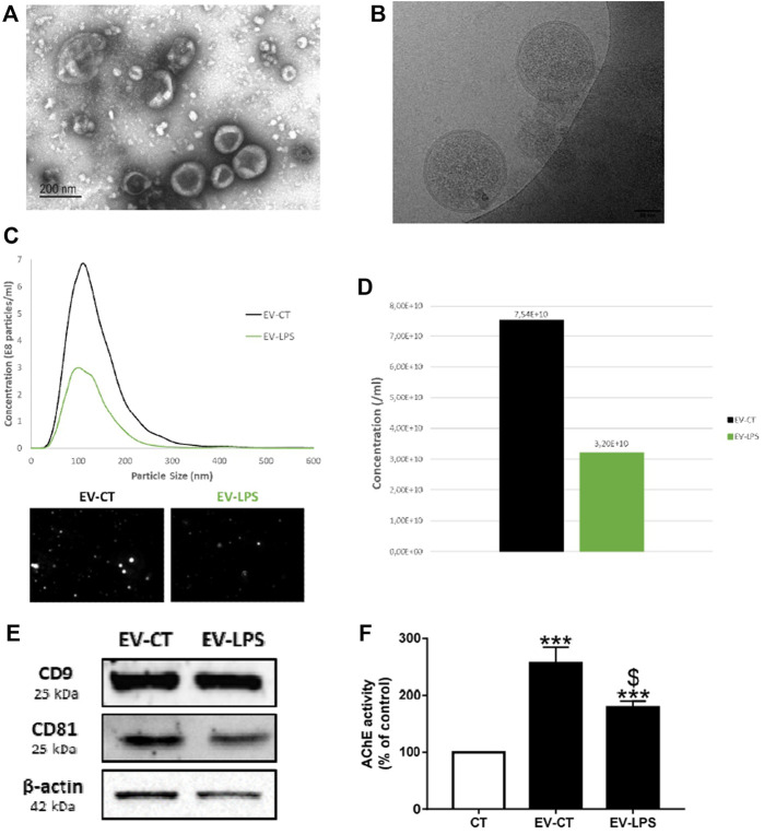
Characterization of macrophage-derived EVs. EVs were isolated from the culture media of RAW cells incubated with (EV-LPS) or without (EV-CT) 1 µg/ml lipopolysaccharide-EK (LPS-EK) for 6 h. Evaluation of EV morphology by (A) transmission electron microscopy (TEM) and (B) cryo-electron microscopy (cryo-EM). (C) Particle-size distribution and (D) the total concentration of macrophage-derived EVs were measured by nanoparticle-tracking analysis (NTA). (E) CD9, CD81, and β-actin protein expression were assessed by western blotting. (F) Quantification of the enzymatic activity of acetylcholinesterase (AChE) in macrophage-derived EVs. Control AChE activity in the assay diluent 1X-D-PBS (CT) was defined as 100%. Data are expressed as the mean ± SEM of seven independent experiments performed in triplicate (n = 7). ***p < 0.001 vs. CT; $p < 0.05 vs. EV-CT, Mann-Whitney test.

