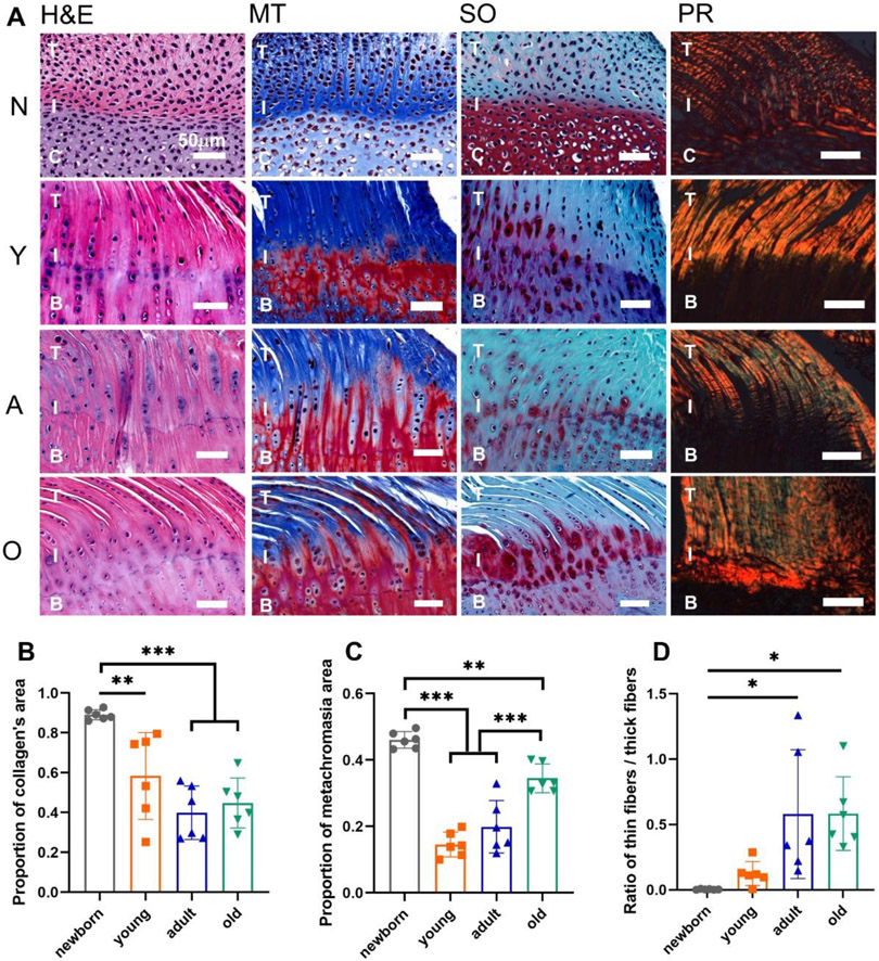
Figure 2.
The histological staining and semi-quantification of the supraspinatus tendon-to-bone interface in four age groups. (A) The H&E, Masson’s Trichrome (MT), Safranin O (SO), and Picrosirius red (PR) staining of the supraspinatus tendon-to-bone interface with the newborn (N), young (Y), adult (A), old (O) groups. (B) The proportion of collagen’s area. (C) The proportion of metachromasia area. (D) The ratio of thin collagen fibers / thick collagen fibers. (T: tendon; C: cartilage; I: interface; B: bone; scale bar: 50 μm; n = 6, *p < 0.05, **p < 0.01, ***p < 0.001)

